This page is general information, not medical advice. Your own doctor or care team is the right source for questions about your skin, diagnosis or treatment.
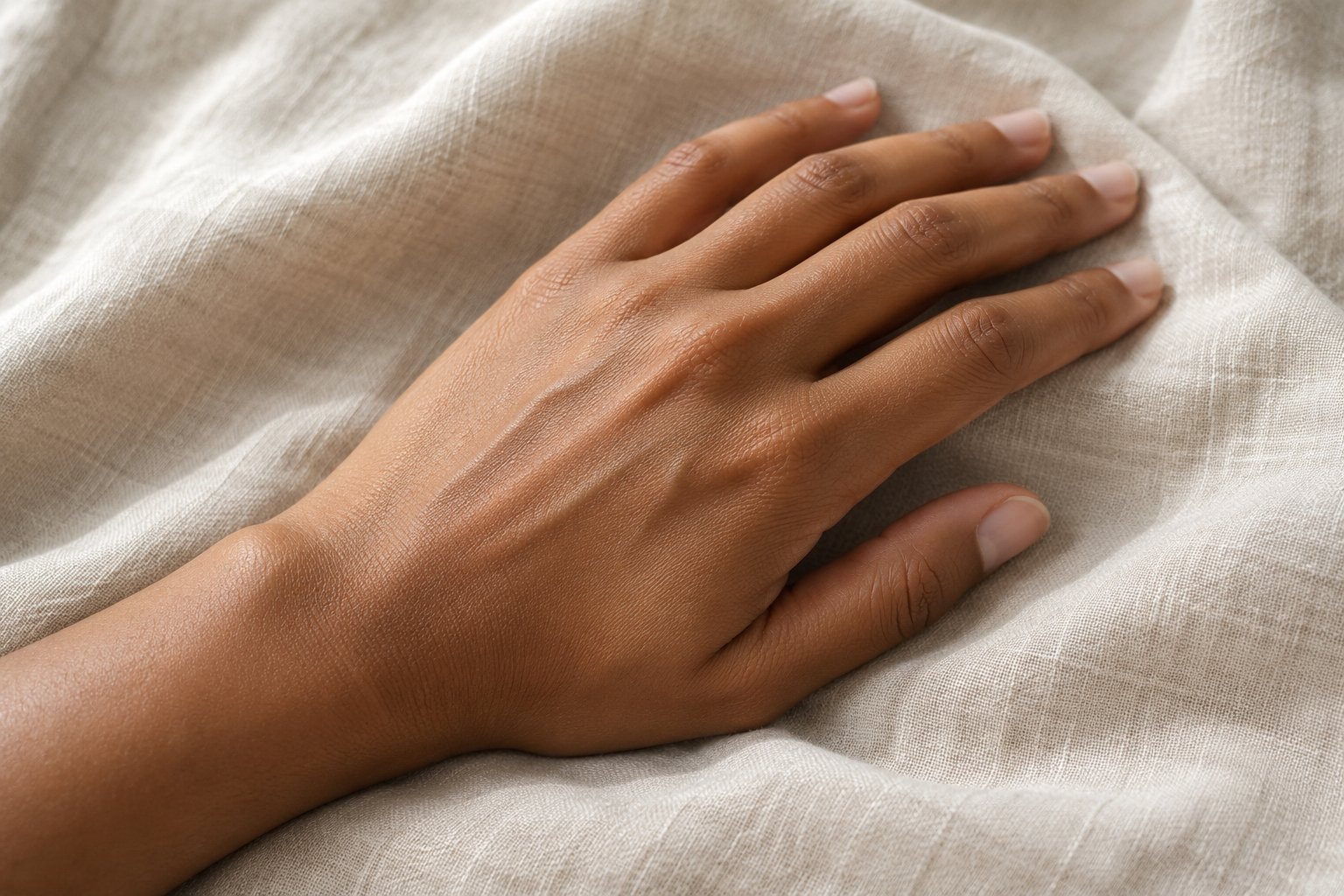
Skin is an organ with a surface, living cells, a supporting network, and a blood supply. It forms a boundary between the body and its surroundings while also sensing contact and helping regulate temperature. Its familiar appearance covers a complicated structure in which cells and the material around them have different jobs.
An organ with many jobs
NIAMS's lesson about skin describes it as the body's largest organ. Its explanation includes a protective covering, a barrier against harmful germs, a role in keeping fluid inside the body, and a sense of touch. The skin also helps keep body temperature within its usual range. These functions depend on more than the layer visible from outside.
The lesson describes nerves that convey information about texture, pressure, and temperature, and sweat-producing cells that help cool the body. Blood vessels, nerves, glands, pigment cells, and supporting fibers are therefore parts of the same organ. Describing the skin as a covering describes only part of its work.
NIAMS also describes hair follicles, the structures from which hair grows, and immune cells within the skin. The sense of touch comes from nerve cells sending messages toward the brain. Different structures are involved in sensing contact, regulating temperature, and maintaining a boundary. They work within tissue that includes both living cells and the material surrounding those cells.
Layers from the surface inward
The epidermis is the outer layer. NIAMS explains that cells form toward its bottom and replace older cells shed from the surface. Below it lies the dermis, with blood vessels, nerve endings, and oil and sweat glands. The tissue farther underneath contains fat and helps store energy.
MedlinePlus's explanation of skin structure and aging describes the dermis as supplying nutrients to the epidermis. It also describes the fat beneath the dermis as padding and insulation. The diagram shows the outer epidermis, the dermis, and the fat beneath, along with a pigment cell and supporting collagen fibers.
The boundaries distinguish tissue with different functions. The surface layer provides the outer barrier, the dermis supplies support and houses vessels and glands, and the fat beneath contributes padding. A view of the surface alone cannot show that entire arrangement.
Surface cells and pigment
Keratinocytes are the main cell type of the epidermis. Wikipedia's keratinocyte reference describes their contribution to the skin barrier. Cells originating in the lower epidermis move outward and change as they go. The outer material is continually shed and replaced, so the skin surface is part of an ongoing cellular process.
Melanocytes are pigment-making cells near the bottom of the epidermis. Wikipedia's melanocyte reference explains that they produce melanin and pass pigment-containing structures to neighboring keratinocytes. Pigment and the cells that make it are different things: melanin is the colored material, while a melanocyte is a living cell.
NIAMS explains that melanin absorbs some of the sun's harmful rays but does not provide complete protection. This gives pigment a role in the organ's response to sunlight without making the amount of pigment a measure of protection from every effect of exposure.
The dermal scaffold
The dermis contains collagen, a structural protein, and elastin, which contributes elasticity. MedlinePlus describes collagen fibers as providing support and elastin fibers as providing flexibility and strength. Wikipedia's elastin reference explains how this protein contributes to a tissue's return toward its original shape after stretching.
These fibers are part of the extracellular matrix, often shortened to ECM: material outside and between cells. Wikipedia's matrix reference describes a network that supplies both structural support and a setting for cell communication. The matrix is a mixture of components with different physical properties rather than a single substance.
Fibroblasts are connective-tissue cells involved in producing that network. Wikipedia's fibroblast reference describes their production of collagen and elastic fibers. The cells and the matrix are closely related: the cells supply matrix components, and the matrix contributes to the physical surroundings in which the cells function.
The extracellular matrix reference describes components being made within resident cells, released into the surrounding space, and assembled with the network already present. Collagen forms fibers within that network. Other components contribute different physical properties. The structure is therefore shaped by how the material is made and organized, as well as by which cells and proteins are present. This is part of the normal biology of connective tissue.
What research reports about the matrix and age
A mini-review published in Gerontology in 2015 described progressive loss and fragmentation of dermal collagen fibers during aging. It examined cellular mechanisms involved in those changes and described their contribution to a thinner, structurally weaker tissue environment. The review concerns how the organ changes, rather than the effect of a product or a procedure.
A review published in 2023 examined skin aging from the perspective of dermal fibroblasts and their relationship with the extracellular matrix. It describes fibroblasts attaching to the matrix and responding to its mechanical properties, meaning the physical forces and structure around them. It also describes changes within the cells themselves.
The review reports a feedback process in which matrix breakdown alters physical forces, changes fibroblast behavior, and contributes to further matrix deterioration. This is a description of interacting cellular and tissue processes. The account considers the organ’s interconnected tissue structure and cells together.
The overlapping work of wound repair
A cut or scrape interrupts the skin's barrier. NIAMS describes blood cells called platelets gathering at an injury to form a clot. The clot helps contain the early response, while cells participate in covering the damaged area. A dried surface clot is a scab, which is different from the new tissue developing beneath it.
Wikipedia's wound-healing reference describes overlapping phases of clotting, inflammation, new tissue formation, and remodeling. Clotting helps close a break in a blood vessel. Inflammation is the response in which cells remove damaged material and other debris. These activities overlap with the work of cells forming new tissue.
During new tissue formation, fibroblasts contribute a temporary matrix and collagen, vessels develop, and surface cells move over the wound area. Remodeling describes later changes in the arrangement of collagen and the removal of cells no longer needed. The reference emphasizes overlap among phases rather than an exact switch from one finished phase to another.
A review published in Nature in 2008 described wound repair as the synchronization of multiple biological pathways after injury. It reported that adult human wound repair commonly results in scar tissue. Its explanation concerns the coordination of biological processes and the tissue formed, rather than instructions for managing a wound.
The passage of time and years of sunlight
Chronological aging describes changes associated with the passage of time. Photoaging describes changes associated with cumulative sunlight exposure. A review published in 2002 described both processes as cumulative and reported that they share fundamental molecular pathways. It distinguished the passage of time from environmental exposure while examining where the processes overlap.
MedlinePlus describes an aging epidermis becoming thinner, changes in connective tissue affecting strength and elasticity, and thinning of the fat beneath the dermis. It also notes changes in glands and blood vessels. These observations concern different parts of the organ rather than only the appearance of wrinkles.
MedlinePlus also describes declining sweat production and oil production with age. Sweat participates in temperature control, while changes in oil production can contribute to dryness. The fat layer’s thinning changes padding and insulation. These observations illustrate how aging can affect several functions of the organ, beyond the supporting fibers alone.
Wikipedia's photoaging reference describes ultraviolet-associated processes involved in breakdown of matrix proteins, including collagen and elastin. That account gives a connection between sunlight exposure and the supporting tissue.
The prevention evidence page considers ultraviolet radiation and skin cancer. The sunscreen and clothing-label explanation describes agency sun-protection terms, and the melanoma basics page explains the disease that begins in pigment-making cells.