This page is general information, not medical advice. Your own doctor or care team is the right source for questions about your skin, diagnosis or treatment.
A suspicious spot can look different from the skin around it, but appearance alone may not tell a doctor exactly what it is. A biopsy is a procedure that removes tissue for examination. For a skin biopsy, the sample comes from the skin. It can be tested for cancer and for other skin conditions.
The tissue examination answers a different question from looking at the skin or making a picture of the inside of the body. It lets a specialist examine the cells themselves. The result describes the sampled tissue, and your own care team explains what that finding means in the wider clinical picture.
Why the tissue sample matters
The National Cancer Institute's explanation of cancer diagnosis says that a biopsy is often the only way to establish cancer with certainty. A pathologist examines the tissue under a microscope and may perform other tests on its cells. The findings are written in a pathology report.
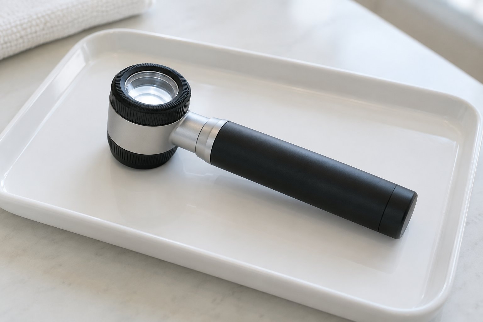
The American Cancer Society's melanoma information explains that some abnormal skin areas are hard to classify by sight. A dermatologist, a doctor specializing in skin disease, may use a lighted magnifying instrument to look more closely. This examination is called dermatoscopy. It helps assess a spot; it does not replace the tissue examination that establishes the diagnosis.
The Society also explains that blood tests are not used to diagnose melanoma. Imaging and other tests may answer questions after melanoma has been found, such as whether it has spread. The distinction between diagnosis and staging is covered in understanding the first weeks after diagnosis.
The main ways a skin sample is taken
MedlinePlus describes skin biopsy methods that include shave, punch and excisional biopsy. The size and location of the abnormal area affect the method used. The American Cancer Society also describes incisional biopsy, which samples part of an abnormal area. The terms tell you how tissue is removed, rather than what the result will be.
- Shave biopsy
- A blade removes tissue from the skin. A superficial shave takes surface layers; a deeper shave includes tissue farther down. The depth matters when the laboratory needs to assess how far an abnormal area extends.
- Punch biopsy
- A round cutting tool removes a small cylinder of tissue. It can include deeper skin layers. The edges of the site may be closed with stitches.
- Excisional biopsy
- The abnormal area is removed in full, usually with a little surrounding skin. The tissue is then examined in the laboratory.
- Incisional biopsy
- Part of the abnormal area is removed for examination. The American Cancer Society describes this when removing the whole suspicious area easily is not possible.
The American Cancer Society's guide to melanoma tests emphasizes that the sample must include enough tissue for an accurate diagnosis. For a suspected melanoma, tissue depth also affects whether the pathologist can measure how deeply it extends into the skin. This explains why the doctor considers more than the visible width of a spot when discussing the procedure.
These descriptions are not a way to select a biopsy method yourself. A doctor can explain the method proposed for a particular area and the information it is expected to provide. A biopsy name does not indicate that melanoma has already been found.
During and after the procedure
MedlinePlus describes cleaning the area and using local numbing medicine before a skin biopsy. Many skin biopsies take place in a doctor's office or another outpatient setting. Depending on the method, the area may need stitches and a dressing. The procedure description can help make the terms familiar, but the exact arrangements come from the clinician performing it.
MedlinePlus lists possible soreness, bruising and bleeding at the biopsy site. The American Cancer Society notes that a biopsy may leave a scar and that different methods can leave different kinds. The person performing the biopsy can explain the expected appearance of the site and the risks relevant to that procedure.
Instructions about dressings, cleaning, stitches and changes at the site are individual clinical instructions. This page does not give a care schedule or explain how to manage a complication. The care team is the source for those details, including which changes they want to hear about and how results will be communicated.
What happens in the laboratory
The National Cancer Institute's pathology-report fact sheet describes tissue being made firm, cut into thin sections and placed on slides. Stains make cell structures easier to see under a microscope. A pathologist reviews the tissue and records a description and diagnosis.
For skin samples, the American Cancer Society explains that a dermatopathologist may read the material. A dermatopathologist has special training in examining skin tissue. Additional laboratory tests may be used when the appearance of the cells does not settle the diagnosis.
A report may describe the sample, microscopic findings and the final diagnosis. If melanoma is identified, features such as tumor thickness become relevant to staging. The page on reading a melanoma pathology report explains that vocabulary and how separate findings fit into a written report. A result seen in a patient portal can appear before the doctor has discussed it; the NCI identifies the treating doctor as the right person to explain it.