Key points
- A skin self-exam is a way to notice changes worth showing a doctor.
- The ABCDE warning signs describe features of a spot; they cannot rule melanoma out.
- A new, changing, itching, bleeding, or unusual spot matters even if it does not match every letter.
- Population screening research has limits, and a visual check is different from a diagnosis.
This page is general information, not medical advice. Your own doctor or care team is the right source for questions about your skin, diagnosis or treatment.
Checking the skin means looking for changes in spots, moles, and other areas. A mole is a cluster of pigment-making skin cells. The purpose of looking is to notice something that deserves medical attention, rather than to decide whether it is cancer. A familiar-looking spot, a small spot, or a spot that has only some warning signs cannot be judged from a checklist alone.
A spot that is new, changing, or different from your other moles is worth showing promptly to a doctor or dermatologist, a doctor who specializes in skin, as the American Academy of Dermatology's self-exam guidance describes. The academy also names itching and bleeding as reasons for a dermatologist to examine a spot.
How the academy describes a skin self-exam
The American Academy of Dermatology describes a sequence for looking over the body, including areas that are difficult to see directly. It uses a full-length mirror and a hand mirror. The following outline explains its sequence. The mirrors give a view of different surfaces; they do not turn the examination into a diagnostic test.
- In a full-length mirror, the academy begins with the front and back of the body. It then describes looking at either side with the arms lifted.
- With the elbows bent, its sequence includes the forearms, underarms, palms, and fingernails.
- It next includes the backs of the legs and feet, the skin between the toes, the toenails, and the soles.
- A hand mirror gives a view of the back of the neck and the scalp. Parting the hair allows a closer look at the scalp itself.
- The final areas in its sequence are the back and buttocks, again viewed with a hand mirror.
This whole-body approach includes places that do not usually face the sun and places easily missed when looking only at the arms or face. The academy's concern is a spot that changes or stands out from the others. Its sequence is a description of where to look, not a statement that a person can exclude cancer after finishing the list.
The ABCDE warning signs
The American Academy of Dermatology's ABCDE reference uses letters to describe warning signs in a mole or pigmented spot. The wording in the table is the academy's. Diameter refers to the width across a spot; evolving refers to change. These are reminders about appearance, rather than requirements that every melanoma must meet.
| Letter | Feature | The academy's description |
|---|---|---|
| A | Asymmetry | One half of the spot is unlike the other half. |
| B | Border | The spot has an irregular, scalloped, or poorly defined border. |
| C | Color | The spot has varying colors from one area to the next, such as shades of tan, brown or black, or areas of white, red, or blue. |
| D | Diameter | While melanomas are usually greater than 6 millimeters, or about the size of a pencil eraser, when diagnosed, they can be smaller. |
| E | Evolving | The spot looks different from the rest or is changing in size, shape, or color. |
The diameter description includes its own limit: melanoma can be smaller. The evolving description also includes a spot that differs from the rest, even before there is a known record of change. A skin check cannot become a pass-or-fail exercise based on how many letters seem to fit. The same concern about a new or unusual spot applies outside the table.
Ordinary moles and other changes
The National Cancer Institute's moles reference distinguishes an ordinary mole from an atypical mole, also called a dysplastic nevus. Ordinary moles are often round or oval, with an even color and a defined edge. An atypical mole can have a less even color, an irregular edge, or a different surface. Neither term, by itself, means melanoma.
The institute explains that melanoma can develop in a mole but more often begins on skin that looked normal. Its warning signs extend beyond color and border: a mole may change in texture or height, become dry or scaly, feel hard or lumpy, itch, or bleed or ooze. These descriptions give language for reporting a change. They do not show what caused it.
The types of melanoma page gives context for the different names used for skin, eye, and mucosal melanomas. The basics page explains the pigment cells from which melanoma begins. Neither an image nor a list of typical appearances can establish what a particular spot is.
A doctor's examination and a tissue diagnosis
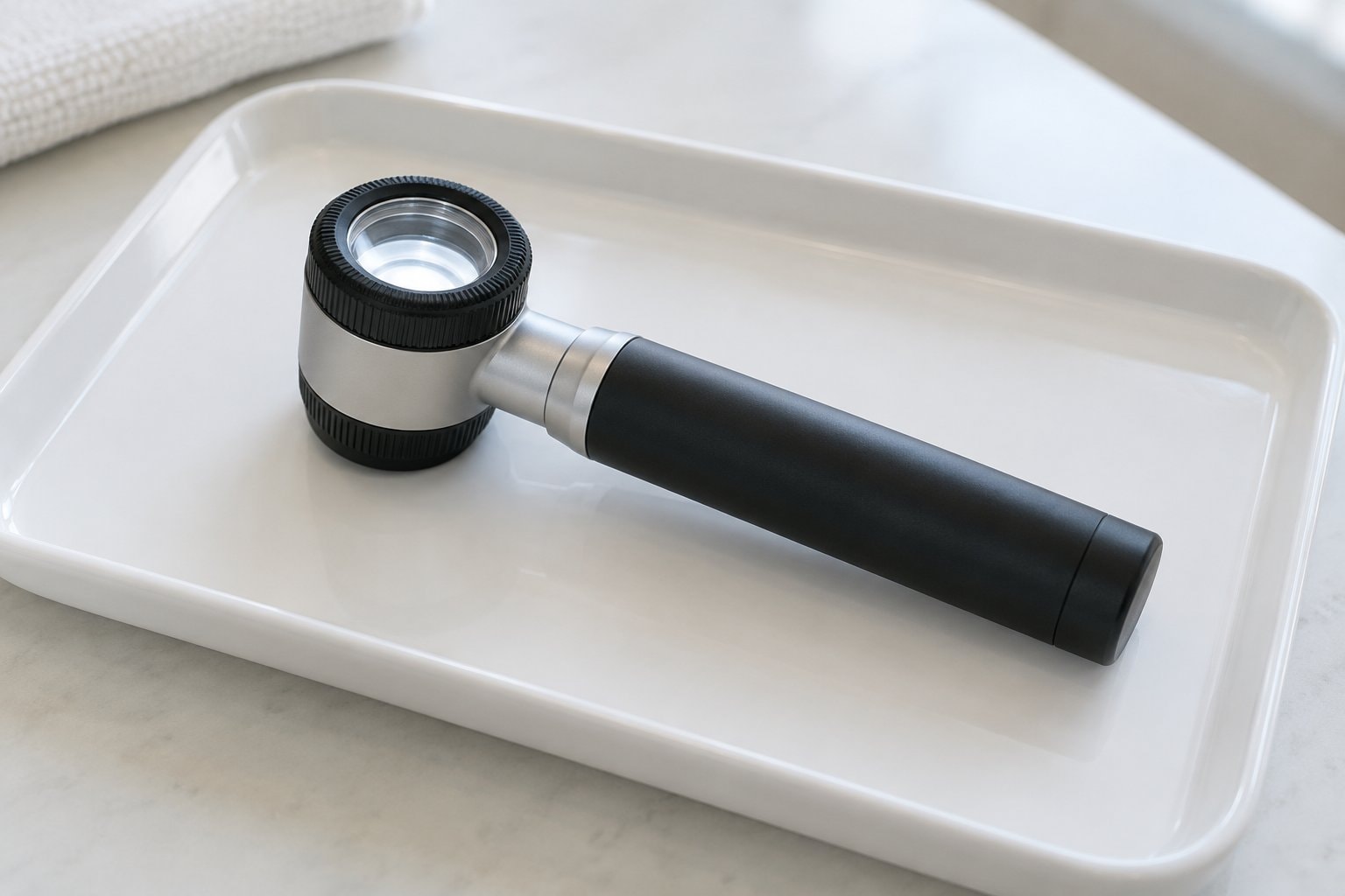
A doctor may use dermatoscopy, also called dermoscopy, to inspect a skin spot. This means examining it with a magnifying instrument and a light source. The instrument helps a clinician see features of the skin; it does not replace examination of a tissue sample when a diagnosis is needed.
The National Cancer Institute says that melanoma is diagnosed by removing tissue and examining it for cancer cells. A biopsy is the removal of that sample. A pathology report records the findings from its examination. The skin biopsy page explains this distinction between looking at a spot and looking at tissue.
What screening evidence can show
The National Cancer Institute's screening summary defines screening as looking for cancer in people without symptoms. It reports that evidence is insufficient to know whether screening the population for skin cancer lowers deaths from the disease. That research question differs from the examination of a specific new or changing spot.
The summary also explains possible false-negative results, when a check appears normal despite cancer, and false-positive results, when a finding looks abnormal but is not cancer. Further tests can have harms, including scarring or infection after a biopsy. These limits are why a self-exam and the ABCDE signs are tools for noticing concerns, rather than a way to rule melanoma out or settle a diagnosis.